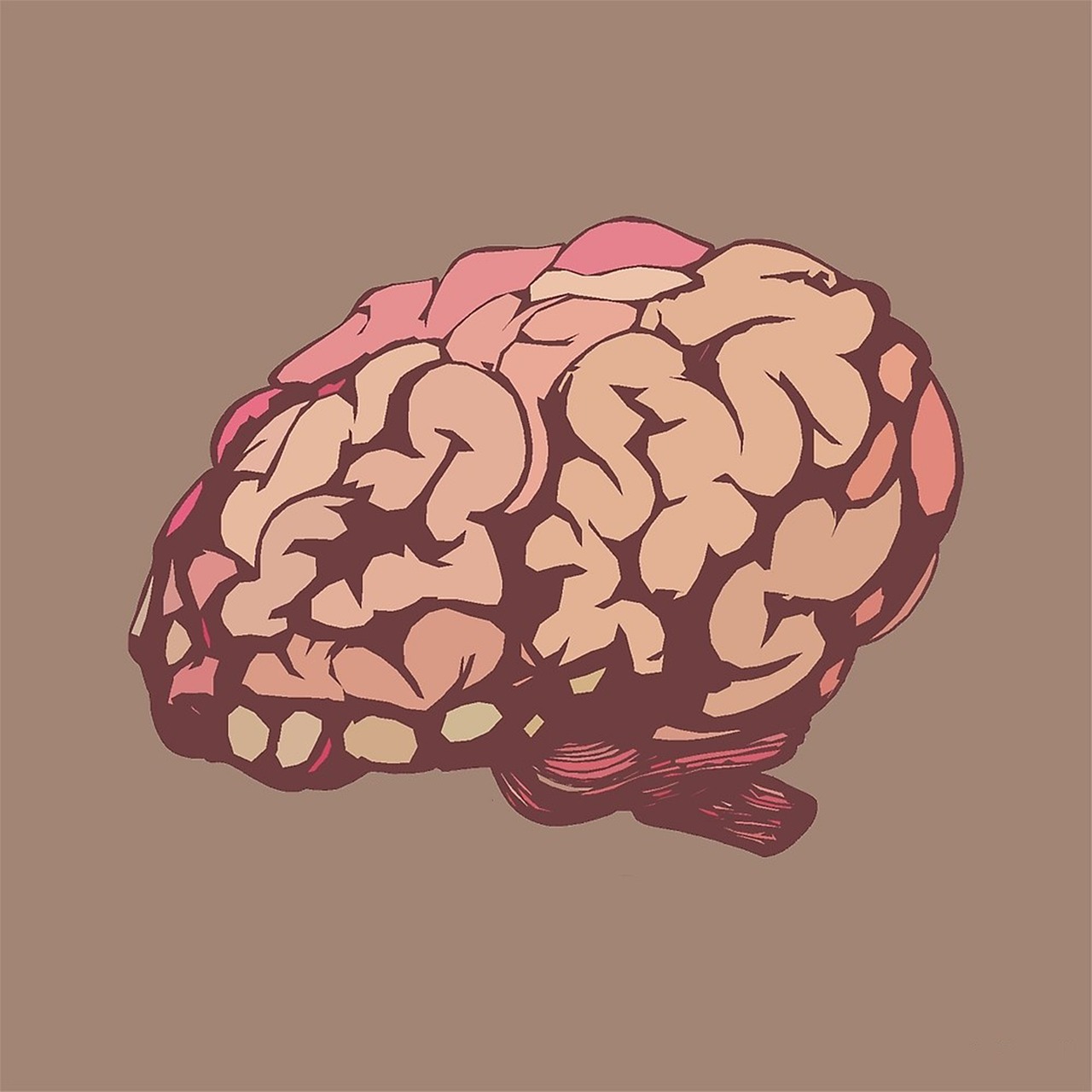
The human brain, with its intricate network of billions of neurons, has long captivated scientists seeking to understand consciousness, memory, and cognition.
Recent breakthroughs in neural microstructure analysis are transforming our ability to visualize and comprehend the brain’s microscopic architecture. These revolutionary advances promise not only to unlock fundamental mysteries of human cognition but also to revolutionize treatments for neurological disorders, enhance artificial intelligence development, and ultimately create a smarter, healthier tomorrow for humanity.
🧠 The New Frontier: Understanding Neural Microstructure
Neural microstructure refers to the microscopic organization of brain tissue, including individual neurons, synapses, glial cells, and the complex connections that form the foundation of all brain activity. For decades, our understanding of this intricate architecture was limited by technological constraints that prevented researchers from observing these structures in living tissue with sufficient detail.
Traditional imaging techniques like standard MRI scans provided valuable information about brain structure but lacked the resolution needed to examine individual neurons and their connections. Electron microscopy offered incredible detail but required brain tissue samples, making it impossible to study living brains in real-time. This gap between macro-level imaging and cellular-level observation left significant blind spots in neuroscience research.
Today’s cutting-edge technologies are bridging this gap, offering unprecedented views into the living brain’s microarchitecture. These advances combine novel imaging modalities, computational algorithms, and artificial intelligence to create detailed maps of neural tissue at resolutions previously thought impossible.
Revolutionary Technologies Transforming Brain Imaging
Diffusion Tensor Imaging and Beyond 🔬
Diffusion Tensor Imaging (DTI) represents one of the most significant advances in non-invasive brain microstructure analysis. This technique measures the directional movement of water molecules along neural pathways, revealing the orientation and integrity of white matter tracts that connect different brain regions.
Building on DTI’s foundation, newer techniques like Neurite Orientation Dispersion and Density Imaging (NODDI) provide even more specific information about neural tissue composition. NODDI can distinguish between different microstructural components, including neurite density, orientation dispersion, and free water content, offering researchers a more nuanced understanding of tissue organization.
These advanced diffusion imaging methods have already demonstrated clinical value in detecting subtle brain changes associated with Alzheimer’s disease, traumatic brain injury, and various psychiatric disorders long before conventional imaging reveals abnormalities.
Connectomics: Mapping the Brain’s Wiring Diagram
Connectomics represents an ambitious effort to create comprehensive maps of neural connections throughout the entire brain. The Human Connectome Project, launched in 2009, has produced increasingly detailed charts of the brain’s structural and functional connectivity patterns.
Recent advances in connectomics combine high-resolution imaging with sophisticated computational analysis to trace individual neural pathways across the brain. These connectivity maps reveal how different brain regions communicate, how information flows through neural networks, and how disruptions in connectivity contribute to neurological and psychiatric conditions.
Machine learning algorithms now analyze connectome data to identify patterns associated with specific cognitive functions, personality traits, and disease states. This information is proving invaluable for developing personalized treatment approaches and predicting individual responses to various interventions.
Artificial Intelligence: The Game-Changer in Neural Analysis 🤖
Artificial intelligence has emerged as an indispensable tool for analyzing the massive datasets generated by advanced brain imaging techniques. Modern neural microstructure studies produce terabytes of data that would be impossible for human researchers to analyze manually.
Deep learning algorithms excel at identifying subtle patterns in brain imaging data that might escape human observation. These AI systems can automatically segment brain structures, trace neural pathways, detect abnormalities, and even predict disease progression based on microstructural changes.
One particularly exciting development involves using generative adversarial networks (GANs) to enhance image resolution beyond the physical limitations of scanning equipment. These algorithms can infer fine details about neural microstructure by learning patterns from high-resolution training datasets, effectively creating “super-resolution” images from standard scans.
Automated Diagnosis and Prognosis
AI-powered analysis of neural microstructure is revolutionizing clinical diagnostics. Machine learning models trained on thousands of brain scans can now identify disease-specific patterns with accuracy matching or exceeding experienced radiologists.
These systems don’t just detect current abnormalities; they can predict future disease development by identifying subtle microstructural changes that precede clinical symptoms. This predictive capability opens unprecedented opportunities for preventive interventions that could halt or slow disease progression before irreversible damage occurs.
Clinical Applications: From Research to Patient Care 🏥
Neurological Disorders and Early Detection
Neural microstructure analysis is transforming how clinicians diagnose and manage neurological disorders. In Alzheimer’s disease research, microstructural imaging can detect changes in brain tissue years before memory symptoms appear, potentially enabling early interventions that preserve cognitive function.
For multiple sclerosis patients, advanced diffusion imaging reveals damage to myelin sheaths surrounding nerve fibers with exceptional sensitivity. This information helps clinicians monitor disease activity, evaluate treatment effectiveness, and adjust therapeutic strategies to optimize outcomes.
Traumatic brain injury research has particularly benefited from microstructural analysis techniques. While standard imaging often appears normal after mild concussions, advanced techniques reveal microscopic disruptions to neural tissue that correlate with cognitive symptoms and predict recovery trajectories.
Psychiatric Conditions and Mental Health
Mental health research is experiencing a paradigm shift as neural microstructure analysis reveals biological underpinnings of psychiatric conditions previously understood primarily through behavioral symptoms. Studies have identified specific microstructural patterns associated with depression, anxiety disorders, schizophrenia, and autism spectrum disorders.
These objective biological markers could transform psychiatric diagnosis from a subjective assessment of symptoms to a more precise, biology-based classification system. Such advances may finally enable truly personalized psychiatric treatment, matching patients to therapies based on their individual brain characteristics rather than trial-and-error approaches.
Brain Development and Aging: A Lifelong Perspective 👶👴
Longitudinal studies using advanced neural microstructure analysis are revealing how the brain develops, matures, and ages at a microscopic level. These insights challenge previous assumptions about brain development and highlight critical windows for interventions.
Research shows that neural microstructure continues evolving well into the third decade of life, particularly in brain regions responsible for executive function and emotional regulation. This extended developmental period has important implications for educational approaches, mental health interventions, and understanding adolescent behavior.
Similarly, studies of healthy aging reveal that some microstructural changes previously attributed to normal aging may actually represent early disease processes. Distinguishing healthy from pathological aging at the microstructural level enables more targeted preventive strategies to maintain cognitive vitality throughout the lifespan.
Enhancing Human Potential: Cognitive Optimization 🚀
Beyond treating disease, neural microstructure research is opening new frontiers in cognitive enhancement and performance optimization. Elite athletes, musicians, and other high performers are increasingly interested in how microstructural brain characteristics relate to exceptional abilities.
Studies comparing experts to novices in various domains reveal specific microstructural adaptations in brain regions critical for their skills. Understanding these neuroplastic changes could inform more effective training methods that accelerate skill acquisition and maximize human potential.
Education and Learning Science
Educational neuroscience is leveraging microstructural brain analysis to understand how learning physically reshapes the brain. These insights are informing evidence-based educational practices optimized for how the brain actually learns and remembers information.
Research has identified microstructural correlates of reading ability, mathematical thinking, and language acquisition. This knowledge helps educators identify students at risk for learning difficulties earlier and develop targeted interventions based on each student’s unique neural profile.
Technical Challenges and Ethical Considerations ⚖️
Data Complexity and Standardization
Despite remarkable progress, neural microstructure analysis faces significant technical challenges. Different imaging protocols, scanner manufacturers, and analysis software can produce varying results, complicating comparisons across studies and clinical sites.
The neuroscience community is actively working toward standardized protocols and quality control measures to ensure reproducibility and reliability. International collaborations are establishing best practices and creating shared databases that enable researchers worldwide to pool data and accelerate discoveries.
Privacy and Ethical Implications
As brain imaging becomes more sophisticated, important ethical questions emerge about privacy, consent, and potential misuse of neurological data. Detailed brain scans could potentially reveal information about personality traits, cognitive abilities, mental health status, and disease predisposition that individuals might prefer to keep private.
Establishing appropriate safeguards while enabling beneficial research requires careful consideration of who should have access to brain imaging data, how it should be stored and protected, and what rights individuals have regarding their own neural information.
The Road Ahead: Future Innovations on the Horizon 🌅
The pace of innovation in neural microstructure analysis shows no signs of slowing. Emerging technologies promise even more dramatic advances in coming years.
Ultra-high field MRI scanners operating at 10.5 Tesla and beyond are pushing the boundaries of in vivo imaging resolution. These powerful magnets enable visualization of brain structures approaching cellular dimensions without requiring invasive procedures.
Optogenetics combined with advanced imaging allows researchers to not only observe neural circuits but also manipulate them with light, testing causal relationships between specific neural pathways and behaviors. These techniques are revealing fundamental principles of how brain circuits encode and process information.
Portable and Accessible Technologies
While cutting-edge brain imaging currently requires expensive equipment in specialized facilities, researchers are developing portable, affordable alternatives that could democratize access to neural analysis. Wearable brain imaging devices using functional near-infrared spectroscopy (fNIRS) and portable MRI systems could bring sophisticated brain monitoring to clinics, schools, and homes worldwide.
These accessibility improvements would enable continuous brain monitoring in natural environments rather than artificial laboratory settings, revealing how neural microstructure and function respond to real-world experiences.
Integration with Other Biological Data 🧬
The future of neural microstructure analysis lies in integration with other biological information sources. Combining brain imaging with genomics, proteomics, metabolomics, and microbiome data creates comprehensive biological profiles that illuminate relationships between brain structure and broader physiological systems.
This systems biology approach reveals how genetic variations, dietary factors, stress responses, and other influences shape individual brain development and function. Such integrated understanding enables truly holistic, personalized approaches to brain health.

Transforming Tomorrow: The Broader Impact 🌍
Revolutionary advances in neural microstructure analysis are catalyzing transformations extending far beyond neuroscience laboratories and medical clinics. These insights are reshaping education, informing artificial intelligence development, inspiring new philosophical perspectives on consciousness, and fundamentally changing how humanity understands itself.
As our ability to visualize and comprehend the brain’s microscopic architecture continues improving, we move closer to answering age-old questions about the nature of thought, emotion, creativity, and consciousness itself. Each discovery raises new questions, ensuring that brain research will remain at the frontier of human knowledge for generations to come.
The journey to unlock the brain’s secrets is far from complete, but the revolutionary tools now available are accelerating progress at an unprecedented pace. From earlier disease detection to cognitive enhancement, from personalized medicine to artificial intelligence inspired by biological neural networks, the implications of understanding neural microstructure touch virtually every aspect of human life.
By continuing to invest in research, addressing ethical challenges thoughtfully, and translating discoveries into practical applications, we are building the foundation for a smarter tomorrow—one where neurological diseases can be prevented or cured, where every individual can reach their cognitive potential, and where the magnificent complexity of the human brain is finally understood in all its intricate glory.