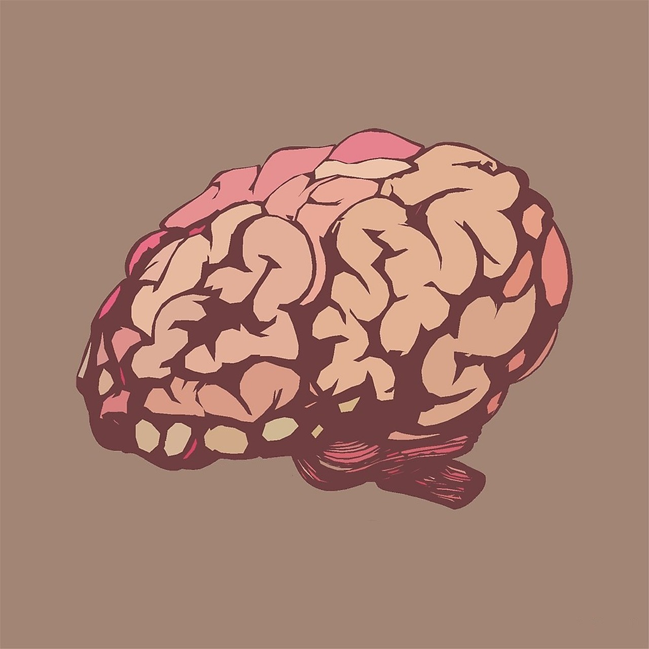
The human brain, with its intricate web of billions of neurons, represents one of the most complex systems in the known universe. Understanding how this remarkable organ processes information, generates thoughts, and controls behavior requires exploring the connections that bind its various regions together.
Modern neuroscience has revolutionized our understanding of the brain by moving beyond studying isolated regions to examining the dynamic networks that enable communication across different neural territories. This shift toward connectivity-based approaches has opened unprecedented windows into both healthy brain function and the mechanisms underlying neurological and psychiatric disorders. By mapping the brain’s structural highways and functional interactions, researchers are decoding the fundamental principles that govern cognition, emotion, and consciousness itself.
🧠 The Foundation: Understanding Brain Connectivity
Brain connectivity refers to the patterns of connections between different neural elements, ranging from individual neurons to large-scale brain regions. This concept has become central to contemporary neuroscience, providing a framework for understanding how distributed brain areas work together to support complex mental processes. Rather than viewing the brain as a collection of specialized modules operating independently, connectivity research reveals an integrated system where information flows continuously across multiple pathways.
The connectivity perspective emerged from advances in neuroimaging technology, computational neuroscience, and network theory. These tools have enabled scientists to visualize and quantify connections that were previously inaccessible to direct observation. The result is a paradigm shift in how we conceptualize brain organization, moving from localizationist views toward more holistic, network-based models of neural function.
Two Complementary Dimensions of Connection
Brain connectivity manifests in two primary forms: structural and functional. Structural connectivity describes the physical pathways—the anatomical “wiring”—that link different brain regions through white matter tracts composed of myelinated axons. These pathways form the substrate through which neural signals can travel, establishing the potential routes for information exchange.
Functional connectivity, in contrast, refers to the statistical relationships between the activity patterns of different brain regions. When two areas show synchronized or correlated activity over time, they are considered functionally connected, even if no direct anatomical pathway links them. This functional coupling can occur through indirect routes involving multiple synaptic relays or through common inputs from other brain structures.
🔬 Mapping Structural Connectivity: The Brain’s Physical Infrastructure
Structural connectivity research aims to chart the complete wiring diagram of the brain—a project known as the connectome. This anatomical blueprint reveals how evolution and development have sculpted the brain’s physical architecture to support efficient information processing. The white matter pathways that constitute structural connectivity vary in size from massive fiber bundles connecting distant brain regions to delicate fascicles linking neighboring areas.
Diffusion tensor imaging (DTI) and its advanced variants represent the primary tools for mapping structural connectivity in living humans. These magnetic resonance imaging techniques exploit the restricted diffusion of water molecules along white matter tracts to reconstruct the three-dimensional trajectories of fiber bundles. By analyzing diffusion patterns across thousands of brain locations, researchers can trace the major pathways connecting different cortical and subcortical structures.
Key Structural Networks in the Human Brain
Several major white matter pathways form the backbone of structural connectivity. The corpus callosum, the brain’s largest commissure, contains approximately 200 million axons connecting the left and right hemispheres, enabling interhemispheric communication and coordination. The arcuate fasciculus links language-related regions in the frontal and temporal lobes, playing a crucial role in speech production and comprehension.
The cingulum bundle follows the curved architecture of the cingulate cortex, connecting frontal, parietal, and temporal regions involved in emotion regulation, memory, and executive control. Association fibers like the superior longitudinal fasciculus and inferior fronto-occipital fasciculus create long-range connections within each hemisphere, supporting integration across sensory, motor, and cognitive domains.
⚡ Functional Connectivity: The Brain in Dynamic Action
While structural connectivity reveals the potential for communication, functional connectivity captures the brain’s moment-to-moment activity patterns and how different regions actually coordinate their operations. Functional connectivity studies typically measure brain activity using functional magnetic resonance imaging (fMRI), which detects blood oxygen level-dependent (BOLD) signals that reflect neural metabolism and activity.
By recording brain activity while participants rest quietly or perform specific tasks, researchers can identify networks of regions that consistently activate together. These synchronized activity patterns suggest that the connected areas are collaborating to support particular mental processes or maintain baseline brain functions.
Resting-State Networks: The Brain’s Default Architecture
One of the most significant discoveries in functional connectivity research is the existence of intrinsic brain networks that persist even when individuals are not engaged in any explicit task. These resting-state networks reveal the brain’s spontaneous organizational principles and baseline functional architecture. The default mode network (DMN) has garnered particular attention, as it shows high activity during wakeful rest and typically deactivates during goal-directed tasks.
The DMN includes key nodes in the medial prefrontal cortex, posterior cingulate cortex, precuneus, and lateral parietal regions. This network is associated with self-referential thinking, autobiographical memory retrieval, future planning, and social cognition. Alterations in DMN connectivity have been linked to numerous conditions including Alzheimer’s disease, depression, and autism spectrum disorders.
Task-Positive Networks and Cognitive Control
In contrast to the default mode network, several task-positive networks show increased activity during demanding cognitive operations. The central executive network, anchored in the dorsolateral prefrontal cortex and posterior parietal cortex, supports working memory, cognitive flexibility, and goal-directed behavior. The salience network, centered on the anterior insula and anterior cingulate cortex, detects behaviorally relevant stimuli and coordinates switches between different network states.
Sensory and motor systems also exhibit strong functional connectivity within their respective domains. Visual networks connect regions throughout the occipital, temporal, and parietal lobes, while motor networks link primary motor cortex with premotor areas, supplementary motor cortex, and the cerebellum. These networks enable coordinated processing within specialized domains while maintaining communication with higher-order cognitive systems.
🔗 The Relationship Between Structure and Function
A fundamental question in neuroscience concerns how structural connectivity constrains and enables functional connectivity. While structural pathways provide the anatomical substrate for communication, the relationship between structure and function is not strictly deterministic. Functionally connected regions do not always share direct structural links, and structurally connected areas may not always show strong functional coupling.
Research indicates that structural connectivity predicts approximately 30-50% of the variance in functional connectivity, suggesting that anatomical pathways provide a foundational framework that is modulated by neural dynamics, synaptic properties, and task demands. Polysynaptic pathways—chains of indirect connections—allow functional communication between regions without direct structural links, explaining how distributed networks can coordinate despite sparse direct anatomical connections.
Network Hubs and Integration
Both structural and functional connectivity analyses reveal that brain networks exhibit non-random organizational principles. Hub regions—areas with disproportionately high connectivity—play crucial roles in integrating information across diverse neural systems. Structural hubs typically include regions like the precuneus, posterior cingulate cortex, and superior frontal cortex, which serve as waypoints for multiple white matter pathways.
Functional hubs often overlap with structural hubs but can also emerge dynamically depending on cognitive context. These highly connected nodes are critical for brain function but also represent vulnerabilities, as their disruption can have cascading effects throughout the network. Many neurological disorders preferentially affect hub regions, potentially explaining why focal damage can produce widespread functional impairments.
📊 Methods and Technologies Advancing Connectivity Research
The explosion of connectivity research reflects technological advances that have made network mapping increasingly feasible and precise. Beyond basic DTI and fMRI, newer techniques offer enhanced resolution and novel perspectives on brain connectivity. High angular resolution diffusion imaging (HARDI) and diffusion spectrum imaging (DSI) can resolve crossing fibers that confound standard DTI, providing more accurate reconstructions of complex white matter architecture.
Magnetoencephalography (MEG) and electroencephalography (EEG) complement fMRI by offering superior temporal resolution, capturing neural dynamics at millisecond timescales. These electrophysiological methods reveal the rapid oscillatory synchronization that underlies functional connectivity, providing insights into the temporal coordination mechanisms that fMRI cannot access.
Graph Theory and Network Analysis
Mathematical frameworks from graph theory have become indispensable tools for quantifying connectivity patterns. By representing the brain as a network of nodes (brain regions) connected by edges (structural or functional connections), researchers can calculate metrics that characterize network topology. Measures like clustering coefficient, path length, modularity, and degree distribution reveal organizational principles such as small-world architecture and modular structure.
Small-world networks balance local clustering with short path lengths between distant nodes, enabling both specialized processing within modules and efficient global integration. The human brain consistently exhibits small-world properties, suggesting evolutionary optimization for information processing efficiency. Network analysis has revealed that many neurological and psychiatric conditions disrupt these optimal organizational principles, shifting network topology toward more random or more regular configurations.
🏥 Clinical Applications: Connectivity in Disease and Disorder
Understanding brain connectivity has profound implications for clinical neuroscience. Virtually every neurological and psychiatric condition involves disruptions to structural or functional connectivity patterns. By characterizing disease-specific connectivity signatures, researchers are developing novel biomarkers for diagnosis, prognosis, and treatment monitoring.
In Alzheimer’s disease, connectivity analysis reveals early disruptions to the default mode network and progressive degradation of white matter pathways, often preceding overt cognitive symptoms. Stroke research uses connectivity mapping to predict recovery outcomes and guide rehabilitation strategies. Traumatic brain injury frequently damages white matter tracts, producing disconnection syndromes where functionally important regions become isolated from their network partners.
Psychiatric Disorders Through the Connectivity Lens
Psychiatric conditions increasingly are viewed as disorders of brain connectivity rather than localized dysfunctions. Schizophrenia shows widespread alterations in both structural and functional connectivity, with particular disruptions in fronto-temporal networks and reduced integration across brain systems. Depression is associated with hyperconnectivity within the default mode network and altered connectivity between emotion-regulating regions.
Autism spectrum disorders exhibit a complex pattern of both over- and under-connectivity, with local overconnectivity in some regions but reduced long-range connectivity between distant brain areas. Attention-deficit/hyperactivity disorder (ADHD) shows immature connectivity patterns, particularly affecting networks involved in cognitive control and attention regulation. These connectivity-based insights are reshaping psychiatric nosology and treatment approaches.
🚀 Future Directions: Toward Dynamic and Personalized Connectivity Maps
The field of connectivity research continues to evolve rapidly, with several emerging directions promising to deepen our understanding. Dynamic functional connectivity—examining how network configurations change over time rather than assuming static patterns—reveals that the brain continuously reconfigures its functional architecture in response to cognitive demands and internal states. These temporal dynamics may prove more informative than static connectivity measures for understanding cognitive flexibility and adaptation.
Personalized connectivity mapping recognizes that individual brains exhibit unique connectivity fingerprints. While group-level patterns reveal general organizational principles, individual differences in connectivity architecture may explain variations in cognitive abilities, personality traits, and disease vulnerability. Precision medicine approaches are beginning to leverage individual connectivity profiles to predict treatment responses and guide personalized interventions.
Integration Across Scales and Modalities
Future connectivity research will increasingly integrate information across multiple spatial and temporal scales. Combining microscopic connectomics—detailed mapping of synaptic connections using electron microscopy—with macroscopic neuroimaging will bridge the gap between cellular neuroscience and systems-level network organization. Multi-modal integration, combining structural, functional, and effective connectivity measures, will provide more complete characterizations of brain network architecture.
Machine learning and artificial intelligence are becoming essential tools for extracting meaningful patterns from complex, high-dimensional connectivity data. These computational approaches can identify subtle network alterations that predict disease trajectories, classify patient subgroups, and discover new connectivity-behavior relationships that might escape traditional statistical methods.

🌟 The Connected Brain: A New Understanding of Mind
Connectivity neuroscience has fundamentally transformed how we conceptualize brain function and dysfunction. Rather than discrete modules performing isolated computations, the brain emerges as a deeply interconnected system where cognition, emotion, and behavior arise from coordinated activity across distributed networks. This network perspective resolves longstanding debates about localization versus distributed processing by showing that both principles operate simultaneously—specialized regions exist, but they function through their embedding within larger networks.
The insights gained from connectivity research extend beyond theoretical neuroscience to practical applications in education, clinical practice, and technology development. Understanding how learning strengthens functional connections can inform educational strategies. Identifying connectivity biomarkers enables earlier diagnosis and more targeted interventions for neurological and psychiatric conditions. Brain-computer interfaces increasingly leverage connectivity patterns to decode intentions and control external devices.
As mapping technologies improve and analytical methods become more sophisticated, our understanding of the brain’s intricate connectivity will continue to deepen. The ultimate goal—a complete, dynamic map of human brain connectivity at multiple scales—remains ambitious but increasingly attainable. This comprehensive understanding promises to unlock fundamental mysteries of consciousness, intelligence, and the nature of the self, while simultaneously providing powerful tools for enhancing brain health and treating disease. The journey to fully understand the brain’s network has only just begun, but the destination promises insights that will reshape our understanding of what it means to be human.